CyberKnife
CyberKnife for brain tumors
Explanation of stereotactic radiosurgery for intracranial tumors and brain metastases: SRS concepts, Gamma Knife differences, lesion number and size, and documents for review.
This information is not a diagnosis or a medical recommendation. Decisions remain with your medical team.
Category, technique, and platform
These three levels are often confused, which can make questions for the doctor less precise. Radiotherapy is the category, SBRT and SRS are techniques, and CyberKnife is one platform that can deliver those techniques.
Radiotherapy
The therapy category: using radiation to control cancer-cell growth in a targeted area.
SBRT and SRS
High-precision stereotactic radiotherapy techniques. SBRT is used for targets outside the head; SRS is used for intracranial targets. Both can be delivered by different platforms.
CyberKnife
One platform that can deliver stereotactic treatment. The platform is robotic and includes motion tracking. It is the tool used, not a separate therapy level.
SRS for intracranial targets
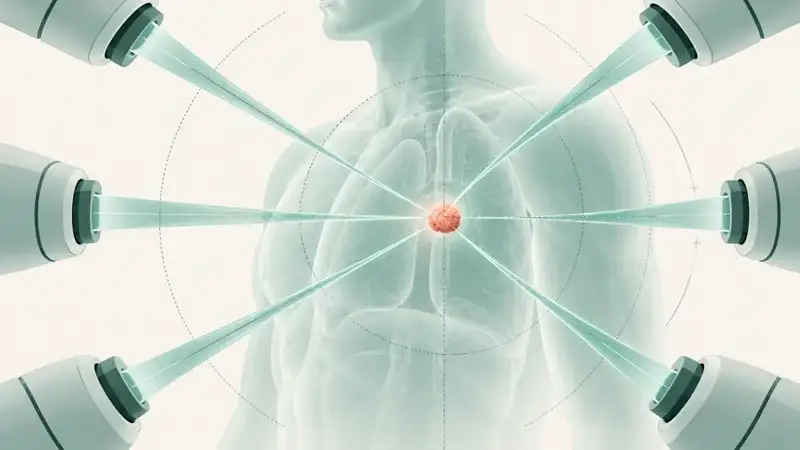
SRS is a stereotactic radiotherapy technique for intracranial targets. The term radiosurgery does not mean surgery is performed: there is no incision and no tissue is removed. Its effect comes from radiation, as with other radiotherapy; the difference is how precisely it is directed.
This technique is commonly discussed for tumors arising inside the head and for brain metastases, which are brain lesions originating from cancer elsewhere in the body.
The relevant question is therefore not whether this is surgery, but whether the target can be clearly defined and the dose can be delivered within safe limits for surrounding tissue.
CyberKnife and Gamma Knife
Both are platform names, not different levels of therapy. Both can deliver stereotactic radiosurgery for targets inside the head.
What usually differs is the workflow, the head-immobilization method, and the range of targets handled at a particular institution. The comparison is institution-dependent: which platform is available, how the team works, and which plan is considered appropriate for an individual case.
TanyaKanker therefore does not conclude which platform is suitable for your case without reviewing the imaging and treatment objective.
Scope of this section
What changes the discussion
Four factors most often change the direction of discussion: the number of lesions, their size, their location inside the head, and any previous radiotherapy. Other treatments already received are also considered. The list below contains inputs for team assessment, not eligibility boxes for a patient to check independently.
Tumor location
How close it is to organs or tissue that must be protected.
Tumor size
Size affects whether focused irradiation can be planned.
Number of lesions
One lesion or several; the number changes the treatment consideration.
Prior radiotherapy
Site, date, dose, and fraction count if any.
Nearby critical organs
Structures that limit the maximum dose that can be delivered.
Motion and breathing
Moving targets change how tracking is used.
Functional status
General condition affects treatment choice.
Treatment goal
Local control, symptom relief, or another agreed goal.
What this list produces
Documents needed for review
For targets in the head, imaging quality determines how far a case can be reviewed. If something is not yet available, send what you already have first.
Most recent CT / MRI / PET-CT
Include DICOM when available; this is often the most important file.
Radiology report
The official reading for the imaging above.
Pathology result
Tumor type and grade when a biopsy has been done.
Prior treatment summary
Surgery, radiotherapy, and systemic therapy with dates.
Prior radiotherapy history
Site, date, dose, fraction count, and plan if accessible.
Molecular / NGS result
When relevant for later options.
Functional status
If known and recorded by the treating team.
When it may not be suitable
This section does not assess an individual case or rule anyone out. It explains why a plan may be judged unsuitable so that your questions for the doctor can be more precise.
- Disease has spread widely so local therapy is no longer the main focus.
- Tumor size or location means focused dose cannot be delivered safely.
- Prior radiotherapy to the same area means cumulative dose limits must be calculated.
- The treatment goal is better answered by another approach, including systemic therapy or surgery.
- The patient's overall clinical condition makes focused therapy a lower priority.
Open frequently asked questions
Frequently asked questions
Short answers to the questions asked most often. The fuller explanation appears in the sections above.
What is different about SRS for targets inside the head?
Intracranial targets require high precision because nearby tissue must be protected from high dose. Head fixation and position verification are important parts of planning.
CyberKnife or Gamma Knife: which is better?
That cannot be answered generally. Both can be used for stereotactic radiosurgery; relevant differences involve workflow, immobilization, and institutional experience.
Does the number of lesions affect therapy choice?
Yes. Number, size, and location change the assessment, including whether focused therapy remains the right approach.
Can it be discussed after previous brain radiotherapy?
It can be discussed for some patients, but it depends on cumulative dose and involved tissue. Prior brain radiotherapy history must be included.
View facts and sources
Source separation
This page separates three things that are often mixed together: provider facts, facts from guidelines or scientific publications, and TanyaKanker explanations. Each line identifies its class so a provider claim is not read as independent evidence.
- PROVIDER
- According to Foshan CyberKnife Center materials
- EXTERNAL
- Guideline or scientific publication
- EXPLAINER
- TanyaKanker explanation
Installed technology
According to Foshan CyberKnife Center materialsCyberKnife M6 (Accuray platform)
Source: Foshan CyberKnife Center materials; consistent with the hospital's official website.
Provider
According to Foshan CyberKnife Center materialsFoshan Fosun Chancheng Hospital, Foshan, Guangdong, China
Source: Official hospital website (www.fsccyy.com).
Facility status
According to Foshan CyberKnife Center materialsPublic JCI-accredited national Grade III-A hospital, operating for almost 60 years
Source: Foshan CyberKnife Center materials.
Technical leadership
According to Foshan CyberKnife Center materialsLed by Dr. Jun Yang, medical physicist; contributor to CyberKnife quality-control standards TG-135 and TG-277
Source: Foshan CyberKnife Center materials.
Contact
According to Foshan CyberKnife Center materialsTel. +86 757 8216 2888 - Address: No.3 Sanyou South Road, Shiwanzhen, Chancheng, Foshan
Source: Foshan CyberKnife Center materials.
Technology origin
Guideline or scientific publicationCyberKnife was developed by Dr. John Adler, professor of neurosurgery at Stanford University; FDA cleared it for tumors in multiple body sites in 2001
Source: Public documentation and regulatory history.
Next step
Build a case summary from your reports so the number and location of lesions and any previous radiotherapy can be reviewed before you decide on the next care step.
This information is not a diagnosis or a medical recommendation. Decisions remain with your medical team.
Explore related pages
Related reading
- CyberKnife overviewCore concepts and the case-review pathway.
- CyberKnife and SBRTUnderstand the category, technique, and platform.
- SRSStereotactic radiosurgery for intracranial targets.
- Brain cancerSubtypes, markers, and treatment options to discuss.
- Clinical trialsA public registry filtered for this disease group.